Anatomical terminology provides a standardized language for describing the human body. It allows healthcare professionals worldwide to communicate clearly. The study of anatomy, from microscopic to macroscopic levels, relies on consistent terms. These terms ensure precise understanding of body structures.
Definition of Anatomy

Anatomy, derived from the Greek words “ana” (up) and “tome” (cutting), is the science that studies the structure of the body. It encompasses the identification and description of the various organs, tissues, and cells that compose the human form. Anatomical study can be approached through different scales, from the microscopic examination of cells (histology) to the macroscopic observation of organs and systems (gross anatomy).
Understanding anatomical structures is crucial for medical professionals. It provides the foundation for diagnosing and treating diseases. Furthermore, anatomy helps in surgical planning and the advancement of medical research. Precise anatomical knowledge is essential for effective healthcare practice.
Importance of Anatomical Terms
Anatomical terms are the cornerstone of medical communication. They provide a precise and universally understood vocabulary for describing the human body. Without standardized terms, ambiguity and misinterpretation can arise, leading to errors in diagnosis and treatment. Anatomical terminology transcends linguistic barriers, enabling healthcare professionals from different countries to collaborate effectively.
Using terms like “superior” and “inferior” instead of “above” and “below” ensures accuracy in describing the relative positions of body parts. Similarly, “anterior” and “posterior” are preferred over “front” and “back”. The consistent application of these terms enhances clarity. Ultimately, anatomical terms improve patient care and safety by reducing the risk of medical errors.

Levels of Anatomical Study
Anatomical study spans different levels of detail, from the microscopic to the macroscopic. These levels offer unique perspectives on the body’s structure and organization. Studying at these levels provides a comprehensive understanding of human anatomy.
Microscopic Anatomy (Histology)
Microscopic anatomy, often called histology, involves studying the body’s tissues and cells using a microscope. This level of anatomical study examines structures too small to be seen with the naked eye. Histology is crucial for understanding the cellular organization of organs and tissues, revealing details about their function and composition. Histological analysis helps identify diseases by examining tissue samples for abnormalities at the cellular level.
The study of histology allows for the observation of cellular structures, such as nuclei, organelles, and cell membranes. Staining techniques enhance the visibility of these components, providing detailed information about tissue types. Microscopic anatomy is essential for diagnosing diseases, researching tissue function, and understanding the body’s overall physiology. Understanding microscopic anatomy is crucial in medical and biological research.
Macroscopic Anatomy (Gross Anatomy)
Macroscopic anatomy, also known as gross anatomy, involves the study of anatomical structures visible to the unaided eye. This branch of anatomy focuses on examining organs, bones, muscles, and other large structures. Gross anatomy provides a foundational understanding of the body’s overall organization and systems. Dissection, imaging techniques, and physical examination are common methods used in gross anatomy studies.
It examines the relationships between different body parts and how they work together. Gross anatomy is essential for medical students, surgeons, and other healthcare professionals. It allows them to visualize the body’s internal structures and understand their functions. This knowledge is crucial for diagnosis, surgical planning, and understanding the impact of injuries and diseases. Gross anatomy provides a comprehensive view of the human body.

Directional Terms
Directional terms are essential for accurately describing the location of structures. These terms, like superior and inferior, provide a common language. Anatomical directions ensure clarity in medical communication. They are vital for precise descriptions.
Superior vs. Inferior
In anatomical terminology, “superior” indicates a position above or higher than another structure relative to the head. Conversely, “inferior” refers to a position below or lower than another structure, closer to the feet. Understanding these terms is crucial for precise anatomical descriptions. For example, the heart is superior to the stomach, while the stomach is inferior to the heart. These directional terms provide a clear frame of reference. This reference is essential for medical professionals. It helps them accurately communicate about the body’s structures. Superior and inferior are fundamental for describing relative positions. Thus, mastering them enhances comprehension in anatomy. These terms are also known as cranial (superior) and caudal (inferior). This knowledge aids in effective communication among healthcare providers and students.
Anterior vs. Posterior
Anterior and posterior are directional terms. They describe the relative position of structures in the body. “Anterior” refers to the front of the body. “Posterior” refers to the back of the body. For example, the sternum is anterior to the heart. The vertebral column is posterior to the heart. These terms are essential for accurately describing anatomical locations. They provide a clear reference point for medical communication. Imagine describing a wound’s location. Using anterior and posterior helps clarify its precise position. These terms are also known as ventral (anterior) and dorsal (posterior). Anatomical descriptions rely heavily on these directional terms. Thus, a strong understanding is critical for healthcare professionals. Mastering anterior and posterior enhances clarity. It ensures effective communication among medical practitioners and students. These terms are indispensable tools in anatomical studies.
Medial vs. Lateral
Medial and lateral are crucial directional terms. They describe a structure’s proximity to the midline of the body. “Medial” indicates closer to the midline. “Lateral” indicates further away from the midline. The nose is medial to the eyes. The ears are lateral to the nose. These terms provide a clear spatial reference. This is essential for accurate anatomical descriptions. Imagine describing the location of a nerve. Medial and lateral help pinpoint its position relative to the body’s center. These terms are fundamental in medical communication. They ensure healthcare professionals understand anatomical relationships correctly. Understanding medial and lateral is vital for anatomical studies. Consider the location of muscles in the limbs. Describing them as medial or lateral clarifies their function. Accuracy is key in anatomical terminology. Medial and lateral contribute to precise descriptions. Mastering these terms enhances comprehension and communication. This is crucial for students and practitioners alike.

Body Planes
Body planes are imaginary flat surfaces. These divide the body into sections. These sections are for anatomical study. The three primary planes are sagittal, coronal, and transverse. Each plane offers a unique perspective. They aid in visualizing internal structures.
Sagittal Plane
The sagittal plane is a vertical plane. It divides the body into right and left parts. If the division is equal, creating mirror images, it’s called midsagittal or median. Parasagittal planes are any sagittal planes offset from the midline. They divide the body into unequal right and left segments. This plane is essential for visualizing internal organs like the brain and spinal cord in a lateral view. Understanding sagittal sections is crucial in medical imaging. It helps doctors identify abnormalities. Anatomical illustrations frequently use sagittal views. These views display relationships between structures. This includes muscles, bones, and organs. Sagittal planes are vital for comprehending the body’s three-dimensional organization. They are also essential for surgical planning and diagnostic procedures.
Coronal (Frontal) Plane
The coronal plane, also known as the frontal plane, is a vertical plane. It divides the body into anterior (front) and posterior (back) parts. Imagine a flat surface slicing through your body from side to side. This division allows viewing structures from the front or back perspective. It’s essential for understanding relationships between muscles, bones, and organs. These relationships are as seen from the anterior or posterior aspect. Medical imaging techniques like CT scans and MRIs frequently use coronal sections. These sections help diagnose medical conditions. Anatomical illustrations often use frontal views. These display the arrangement of organs within the thoracic and abdominal cavities. The coronal plane differs from the sagittal plane, which divides the body into left and right sections. Understanding the coronal plane is vital. It is vital for medical professionals in various fields. It is also important for students studying anatomy.
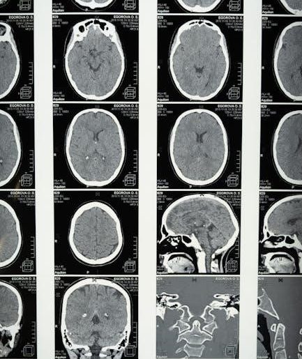
Transverse Plane
The transverse plane, also called the horizontal or axial plane, divides the body into superior (upper) and inferior (lower) sections. Envision a horizontal cut through the torso, separating the head from the feet. This plane is crucial for understanding cross-sectional anatomy. It allows detailed visualization of internal organs and structures. Medical imaging relies heavily on transverse sections. CT scans and MRIs frequently provide images in this plane. These are essential for diagnosing and treating various medical conditions. Anatomical atlases and textbooks often include transverse views. These views enhance understanding of organ placement. These views also show the relationship to surrounding tissues. The transverse plane differs from the sagittal and coronal planes. It offers a unique perspective for studying anatomy. It is particularly useful for assessing abdominal and pelvic anatomy. Mastery of the transverse plane is vital. This mastery is vital for healthcare professionals. It is also important for students studying anatomy and physiology.

Regional Terms
Regional terms are used to specify specific areas of the body. These terms provide clear anatomical location. They are essential for accurately describing anatomical features. Understanding regional terms is crucial in medicine and anatomy studies.
Abdominal Region

The abdominal region is a significant area of the anterior trunk. It is located below the thorax and above the pelvic region. The abdominal region houses many vital organs. These include the stomach, intestines, liver, pancreas, and gallbladder. The abdominal region can be further divided into quadrants or regions for more precise anatomical description. Common terms related to the abdominal region include epigastric, umbilical, and hypogastric. The inguinal region, or groin area, is also closely associated with the abdominal region. Understanding the anatomy of the abdominal region is critical for diagnosing and treating various medical conditions. The abdominal region is a complex area containing many layers of muscles and tissues. Pain or discomfort in the abdominal region can indicate a wide range of health issues. Clinical examination of the abdominal region often involves palpation and auscultation. Knowledge of the abdominal region is essential for medical professionals.

Thoracic Region
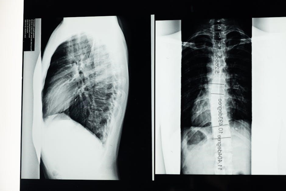
The thoracic region, commonly referred to as the chest, is a vital area of the body. It is located between the neck and the abdominal region. The thoracic region contains several essential organs, including the heart and lungs. The sternum, located in the middle of the chest, is an important landmark. The pectoral region also refers to the chest area, specifically the anterior portion. The thoracic region is protected by the rib cage, which consists of twelve pairs of ribs. The intercostal muscles are located between the ribs, aiding in respiration. The scapular region, or shoulder blade area, is located on the posterior aspect of the thoracic region. The mediastinum, a space within the thoracic cavity, contains the heart, major blood vessels, trachea, and esophagus. Understanding the anatomy of the thoracic region is crucial for diagnosing and treating respiratory and cardiovascular diseases. Imaging techniques, such as X-rays and CT scans, are commonly used to visualize the structures within the thoracic region. The thoracic region is essential for breathing and circulation.
Pelvic Region
The pelvic region, located inferior to the abdominal region, houses several important organs. This region contains the bladder, rectum, and reproductive organs. The pelvic region is bounded by the pelvic bones, forming a basin-like structure. Muscles of the pelvic floor support the pelvic organs. The perineal region, located between the thighs, is part of the pelvic region. The inguinal region, or groin area, is situated at the junction of the thigh and abdomen. The pelvic region is crucial for urinary, digestive, and reproductive functions. Understanding the anatomy of the pelvic region is essential for diagnosing and treating conditions such as pelvic inflammatory disease and prostate cancer. The ilium, part of the hip bone, contributes to the structure of the pelvis. The obturator foramen, a large opening in the pelvic bone, allows passage of nerves and blood vessels. In females, the pelvic region also contains the uterus, ovaries, and fallopian tubes. The pelvic region plays a crucial role in childbirth. The sacrum forms the posterior part of the pelvic girdle.

Anatomical Position
The anatomical position serves as a standard reference point in anatomy. In this position, the body is erect, facing forward, with feet flat on the ground. The arms are at the sides, with palms facing forward. The thumbs point away from the body. This standardized posture ensures consistent descriptions of body parts. All anatomical terms are based on this position, regardless of the body’s actual orientation. This allows for clear communication among healthcare professionals. The anatomical position is crucial for understanding directional terms like superior, inferior, anterior, and posterior. Without a standard reference, these terms would be ambiguous. Imagine trying to describe the location of an organ if the body’s position wasn’t defined. The anatomical position is not necessarily a natural or comfortable posture. It is simply a convention that facilitates accurate anatomical descriptions. When studying anatomy, always visualize the body in this position. This will help you understand the relationships between different body structures. The anatomical position is the foundation of anatomical terminology. It ensures that everyone is on the same page when discussing the human body. It is universally accepted as the starting point for anatomical descriptions.